Every living thing starts with a cell
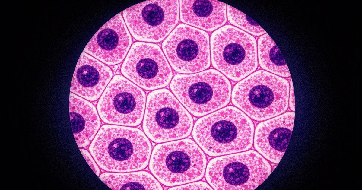
Before you can understand how a heart pumps blood or how a leaf captures sunlight, you need to understand the building block that makes all of it possible: the cell. Every organism on Earth, from a bacterium to a blue whale, is built from cells. Some organisms consist of just one (unicellular), while others are made of trillions (multicellular). For your IGCSE Biology exam, this topic is foundational. Almost every other topic connects back to cell structure in some way, so getting this right pays dividends across the entire syllabus.
Animal cells vs plant cells vs bacterial cells
The first problem to solve is straightforward: what structures does each type of cell contain, and what does each structure do? Think of it as an inventory check. If you can list the parts and match them to their jobs, you have the core of this topic covered.
| Structure | Function | Animal cell | Plant cell | Bacterial cell |
|---|---|---|---|---|
| Cell membrane | Controls what enters and leaves the cell; a selectively permeable barrier | Yes | Yes | Yes |
| Cell wall | Provides structural support and prevents the cell from bursting | No | Yes (cellulose) | Yes (not cellulose) |
| Nucleus | Contains genetic material (DNA) that controls cell activities | Yes | Yes | No (has circular DNA instead) |
| Cytoplasm | Jelly-like substance where most chemical reactions occur | Yes | Yes | Yes |
| Mitochondria | Site of aerobic respiration; releases energy from glucose | Yes | Yes | No |
| Ribosomes | Site of protein synthesis | Yes | Yes | Yes (smaller) |
| Chloroplasts | Contain chlorophyll; site of photosynthesis | No | Yes | No |
| Large permanent vacuole | Filled with cell sap; maintains turgor pressure | No (small temporary ones only) | Yes | No |
| Circular DNA | Carries genetic information (not enclosed in a nucleus) | No | No | Yes |
| Plasmids | Small extra rings of DNA; can carry genes for antibiotic resistance | No | No | Yes |
Reading the table like a problem
Here is how to use that table on exam day. Suppose a question shows you a diagram of a cell and asks: "Is this an animal cell, a plant cell, or a bacterial cell? Explain your answer."
Your method:
- Check for a nucleus. If there is no membrane-bound nucleus, it is a bacterial cell.
- If a nucleus is present, check for a cell wall and chloroplasts. If both are present, it is a plant cell.
- If a nucleus is present but there is no cell wall and no chloroplasts, it is an animal cell.
That three-step decision process will handle any identification question the exam throws at you.
What each structure actually does
Knowing the names is only half the job. Examiners regularly ask "state the function of" a named structure. Here is a concise reference:
- Cell membrane: controls the passage of substances into and out of the cell. It is selectively permeable, meaning it lets some molecules through but blocks others.
- Cell wall: made of cellulose in plants. It is fully permeable (unlike the membrane) and provides rigid support so the cell keeps its shape.
- Nucleus: contains DNA, which carries the instructions for making proteins. It controls the cell's activities.
- Cytoplasm: a watery gel where enzymes catalyse chemical reactions. Most metabolic reactions happen here.
- Mitochondria: the site of aerobic respiration. They break down glucose to release energy for the cell's processes.
- Ribosomes: tiny structures where amino acids are assembled into proteins (protein synthesis).
- Chloroplasts: contain the green pigment chlorophyll, which absorbs light energy for photosynthesis. Only found in green parts of plants.
- Vacuole (plant): a large, permanent, fluid-filled space that stores cell sap (a solution of sugars and salts). It helps maintain turgor pressure, keeping the cell firm.
New cells come from existing cells
This is a short but important syllabus point. New cells are produced by the division of existing cells. A cell grows, copies its DNA, and then splits into two. The IGCSE syllabus does not require you to describe mitosis in detail at Core level, but you must know the principle: cells divide to produce new cells for growth and repair.
Cell specialisation and levels of organisation
A freshly divided cell starts out fairly generic. Through a process called differentiation, it develops specific structures that allow it to carry out a particular function. Once specialised, a cell is adapted for one job and does it well.
Examples of specialised cells
| Specialised cell | Key structural adaptation | Function |
|---|---|---|
| Root hair cell | Long, thin extension (the "hair") that increases surface area | Absorbs water and mineral ions from the soil |
| Red blood cell | Biconcave disc shape (no nucleus in mammals), packed with haemoglobin | Transports oxygen around the body |
| Nerve cell (neurone) | Very long axon, branched endings (dendrites) | Carries electrical impulses rapidly across the body |
| Sperm cell | Streamlined head, long tail (flagellum), many mitochondria | Swims to and fertilises the egg cell |
| Palisade mesophyll cell | Tall, column-shaped, packed with chloroplasts near the upper surface | Carries out most of the leaf's photosynthesis |
| Ciliated epithelial cell | Tiny hair-like projections (cilia) on the surface | Wafts mucus (and trapped particles) along airways |
The logic behind every specialised cell is the same: structure matches function. If you can explain how a cell's shape or contents help it do its job, you can answer any specialisation question.
The hierarchy of organisation (Supplement)
Specialised cells do not work in isolation. They are organised into increasingly complex levels:
- Cell - the basic unit of life
- Tissue - a group of similar cells working together to perform a shared function (e.g. muscle tissue, epithelial tissue)
- Organ - a structure made of different tissues working together (e.g. the heart contains muscle tissue, blood vessel tissue and nerve tissue)
- Organ system - a group of organs working together to carry out a major body function (e.g. the digestive system includes the stomach, intestines, liver and pancreas)
- Organism - the complete living thing, made up of all its organ systems functioning together
Magnification and size calculations
This is the most mathematical part of IGCSE Biology, and it catches students out more than almost any other topic. The good news: there is only one formula, and once you can rearrange it, every question follows the same pattern.
The formula:
Magnification = Image size / Actual size
Or rearranged:
- Actual size = Image size / Magnification
- Image size = Actual size x Magnification
Unit conversions
Before you can plug numbers into the formula, every measurement must be in the same unit. Here are the conversions you need:
- 1 mm = 1000 um (micrometres)
- 1 um = 1000 nm (nanometres)
- 1 mm = 1,000,000 nm
To convert from a larger unit to a smaller one, multiply. To convert from smaller to larger, divide.
Worked example 1: finding magnification
A photograph of a cell measures 30 mm across. The actual cell is 60 um wide. What is the magnification?
Step 1: Convert to the same unit. The image is 30 mm. Convert the actual size to mm: 60 um = 60 / 1000 = 0.06 mm.
Step 2: Apply the formula. Magnification = 30 / 0.06 = x500.
The image is 500 times larger than the real cell.
Worked example 2: finding actual size
A drawing of a bacterium is 45 mm long. The magnification is x1500. What is the actual length?
Step 1: Actual size = Image size / Magnification = 45 / 1500 = 0.03 mm.
Step 2: Convert to micrometres if needed: 0.03 x 1000 = 30 um.
Worked example 3: using a scale bar (Supplement)
A micrograph has a scale bar labelled "10 um" that measures 20 mm on the printed image. A structure in the same image measures 35 mm. What is the actual size of the structure?
Step 1: Find the magnification from the scale bar. The bar represents 10 um = 0.01 mm actual. Image size of bar = 20 mm. Magnification = 20 / 0.01 = x2000.
Step 2: Use the magnification to find the actual size of the structure. Actual size = 35 / 2000 = 0.0175 mm = 17.5 um.
Common exam mistakes
- Confusing cell wall and cell membrane: the wall is rigid and fully permeable (cellulose); the membrane is flexible and selectively permeable. They are not interchangeable terms.
- Saying animal cells have no vacuole: animal cells can have small, temporary vacuoles. What they lack is a large, permanent vacuole. Use the word "permanent" to be precise.
- Forgetting unit conversions in magnification questions: if the image is in mm and the actual size is in um, you must convert before dividing. Skipping this step gives an answer that is 1000 times too large or too small.
- Stating that plant cells have no mitochondria: they do. Plant cells carry out respiration just like animal cells. Chloroplasts are for photosynthesis; mitochondria are for respiration. Both processes happen in plant cells.
- Writing "controls what goes in and out" for the nucleus: that is the cell membrane's job. The nucleus controls cell activities by containing the genetic instructions (DNA).
- Mixing up magnification and resolution: magnification is how much larger the image is than the real object. Resolution is the ability to distinguish between two points that are close together. A blurry image can have high magnification but low resolution.
- Listing "energy" as a structure function without context: mitochondria release energy through respiration. Saying a structure "gives the cell energy" is too vague. State that it is the site of aerobic respiration.
Self-check questions
- Name three structures found in plant cells but not in animal cells.
- A bacterial cell has no nucleus. How does it still carry genetic information?
- A micrograph shows a cell that is 48 mm wide in the image. The magnification is x800. Calculate the actual width of the cell in micrometres.
- Explain how the structure of a root hair cell is adapted to its function.
- Put these in order from simplest to most complex: organ, tissue, organism, cell, organ system.
- A red blood cell has no nucleus. Explain one advantage of this for its function of carrying oxygen.
- Convert 0.25 mm into micrometres and then into nanometres.

Cells are the fundamental units of life, and understanding their structure, function and organisation is essential for IGCSE Biology. This guide covers animal, plant and bacterial cell structures, magnification calculations, unit conversions, cell specialisation and the hierarchy from cells to organ systems, with worked examples, common mistakes and self-check questions.


Comentario(s)